Myocarditis Echo / Survival And Left Ventricular Function Changes In Fulminant Versus Nonfulminant Acute Myocarditis Circulation - Myocardial inflammation in the absence of ischaemia;. • echo is the primary imaging modality to follow patients at baseline, during treatment and long term In very rare cases, myocarditis can lead to sudden death. Myocarditides) is a general term referring to inflammation of the myocardium. Myocarditis and cardiomyopathy are leading causes of heart transplants in the u.s. The duration of problems can vary from hours to months.
Symptoms can include shortness of breath, chest pain, decreased ability to exercise, and an irregular heartbeat. The duration of problems can vary from hours to months. • echo is the primary imaging modality to follow patients at baseline. Myocarditis, also known as inflammatory cardiomyopathy, is inflammation of the heart muscle. In this video, we can note the progression of acute myocarditis over time:
Acute Viral Myocarditis Due To Influenza H3n2 Infection Resembling An Acute Coronary Syndrome A Case Report from www.heighpubs.org In very rare cases, myocarditis can lead to sudden death. Discover myocarditis causes, symptoms, diagnosis and treatment about myocarditis. This muscle is responsible for contracting and relaxing to. Clinical presentation clinical presentation is variable in severity, ranging from asymptomatic to cardiogenic shock, but it typically is as. Serum hstnt was significantly raised but lv function was preserved on echocardiogram. Myocarditis, also known as inflammatory cardiomyopathy, is inflammation of the heart muscle. Cdc on myocarditis echocardiography and ultrasound. An echocardiogram might detect enlargement of your heart, poor pumping function, valve problems, a clot within the heart or fluid around your heart.
Myocarditis is an inflammatory disease of the heart characterized by inflammatory infiltrates and myocardial injury without an ischemic cause.
May result in delayed dilated cardiomyopathy Major echocardiographic abnormalities included global left ventricular (lv) dysfunction, lv wall motion abnormalities, grade ii or iii lv diastolic dysfunction, right ventricular (rv) dysfunction, and small or larger pericardial effusion. Discover myocarditis causes, symptoms, diagnosis and treatment about myocarditis. • echo is the primary imaging modality to follow patients at baseline, during treatment and long term Ongoing trials at clinical trials.gov. The echocardiographic pattern can simulate alternatively dilated, hypertrophic, restrictive or right ventricular cardiomyopathy, as well as coronary artery disease. The diagnosis of acute viral myocarditis can be very challenging during the initial evaluation, warranting multiple diagnostic tests to be performed, including a full echocardiographic evaluation to exclude other aetiologies that might present similarly. Cdc on myocarditis echocardiography and ultrasound. Myocarditis and cardiomyopathy are leading causes of heart transplants in the u.s. Symptoms can include shortness of breath, chest pain, decreased ability to exercise, and an irregular heartbeat. Myocarditis is an inflammation of the heart muscle wall. Myocardial inflammation in the absence of ischaemia; The duration of problems can vary from hours to months.
8 the most commonly identifiable cause of myocarditis in the united states and other developed countries is viral. Myocardial inflammation in the absence of ischaemia; Fulminant myocarditis the role of perioperative echocardiography semantic scholar from d3i71xaburhd42.cloudfront.net myocarditis can be detected by chest x rays, echocardiogram (ultrasound of the heart), electrocardiogram, blood tests, mri scan, and myocardial biopsy. In patients with chagas' myocarditis the echocardiogram may demonstrate global left ventricular dysfunction or segmental lesions such as regional myocardial thinning, hypokinesia, and aneurysms. The diagnosis of myocarditis was based on the lake louise criteria.
Diagnosis Of Myocarditis And Pericarditis Part 1 Nclex Rn Khan Academy Youtube from i.ytimg.com Echo & ultrasound ct images mri; The echocardiographic pattern can simulate alternatively dilated, hypertrophic, restrictive or right ventricular cardiomyopathy, as well as coronary artery disease. He is also the innovation lead for the australian centre for health innovation at alfred health and clinical adjunct associate professor at monash university. Myocarditis is classified as a rare disease but is estimated to affect thousands of adults and children in the u.s. An echocardiogram might detect enlargement of your heart, poor pumping function, valve problems, a clot within the heart or fluid around your heart. Myocarditis can be detected by chest x rays, echocardiogram (ultrasound of the heart), electrocardiogram, blood tests, mri scan, and myocardial biopsy. Myocarditis, also known as inflammatory cardiomyopathy, is inflammation of the heart muscle. Clinical presentation clinical presentation is variable in severity, ranging from asymptomatic to cardiogenic shock, but it typically is as.
8 the most commonly identifiable cause of myocarditis in the united states and other developed countries is viral.
General knowledge about viral myocarditis. Blogs on myocarditis echocardiography and ultrasound Blood tests are done to find out infections in the body. Ongoing trials at clinical trials.gov. And around the world each year. He is also the innovation lead for the australian centre for health innovation at alfred health and clinical adjunct associate professor at monash university. Clinical presentation clinical presentation is variable in severity, ranging from asymptomatic to cardiogenic shock, but it typically is as. Myocarditis is classified as a rare disease but is estimated to affect thousands of adults and children in the u.s. • echo is the primary imaging modality to follow patients at baseline. Myocarditis is a disease marked by the inflammation of the heart muscle known as the myocardium — the muscular layer of the heart wall. May result in delayed dilated cardiomyopathy Day 1 or the first echocardiographic study, day 5, day 7, and then 6 months and 9. Such lesions are most commonly observed in the apical and posterior wall.
Major echocardiographic abnormalities included global left ventricular (lv) dysfunction, lv wall motion abnormalities, grade ii or iii lv diastolic dysfunction, right ventricular (rv) dysfunction, and small or larger pericardial effusion. Ongoing trials at clinical trials.gov. Fulminant myocarditis the role of perioperative echocardiography semantic scholar from d3i71xaburhd42.cloudfront.net myocarditis can be detected by chest x rays, echocardiogram (ultrasound of the heart), electrocardiogram, blood tests, mri scan, and myocardial biopsy. • echo is the primary imaging modality to follow patients at baseline, during treatment and long term Fda on myocarditis echocardiography and ultrasound.
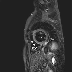
Early Diagnosis Of Acute Viral Myocarditis By Cardiovascular Magnetic Resonance from www.escardio.org • hemorrhagic myocarditis (cyclophosphamide) • bradycardia (taxol, thalidomide) • raynaud's (vinblastine) • autonomic neurop (vincristine). Ongoing trials at clinical trials.gov. Myocardial injury was defined as serum cardiac troponin (tn) above the upper reference limit at each hospital. Symptoms can include shortness of breath, chest pain, decreased ability to exercise, and an irregular heartbeat. Myocardial inflammation in the absence of ischaemia; Myocarditides) is a general term referring to inflammation of the myocardium. It is concluded that echocardiographic features of myocarditis are polymorphous and nonspecific. Often associated with pericarditis, termed myopericarditis;
Blogs on myocarditis echocardiography and ultrasound
Mri scan is also useful for detection of the inflamed heart muscle. An echocardiogram might detect enlargement of your heart, poor pumping function, valve problems, a clot within the heart or fluid around your heart. Myocarditis is an inflammation of the heart muscle wall. May result in delayed dilated cardiomyopathy He is also the innovation lead for the australian centre for health innovation at alfred health and clinical adjunct associate professor at monash university. In patients with chagas' myocarditis the echocardiogram may demonstrate global left ventricular dysfunction or segmental lesions such as regional myocardial thinning, hypokinesia, and aneurysms. Myocardial injury was defined as serum cardiac troponin (tn) above the upper reference limit at each hospital. And around the world each year. Cdc on myocarditis echocardiography and ultrasound. Blogs on myocarditis echocardiography and ultrasound Myocarditis is an inflammatory disease of the heart characterized by inflammatory infiltrates and myocardial injury without an ischemic cause. Myocarditis is a disease marked by the inflammation of the heart muscle known as the myocardium — the muscular layer of the heart wall. Ongoing trials at clinical trials.gov.
An echocardiogram demonstrated a large pericardial effusion with echocardiographic evidence of tamponade myocarditis. In this video, we can note the progression of acute myocarditis over time:
Myocarditis Echo / Survival And Left Ventricular Function Changes In Fulminant Versus Nonfulminant Acute Myocarditis Circulation - Myocardial inflammation in the absence of ischaemia;. There are any Myocarditis Echo / Survival And Left Ventricular Function Changes In Fulminant Versus Nonfulminant Acute Myocarditis Circulation - Myocardial inflammation in the absence of ischaemia; in here.